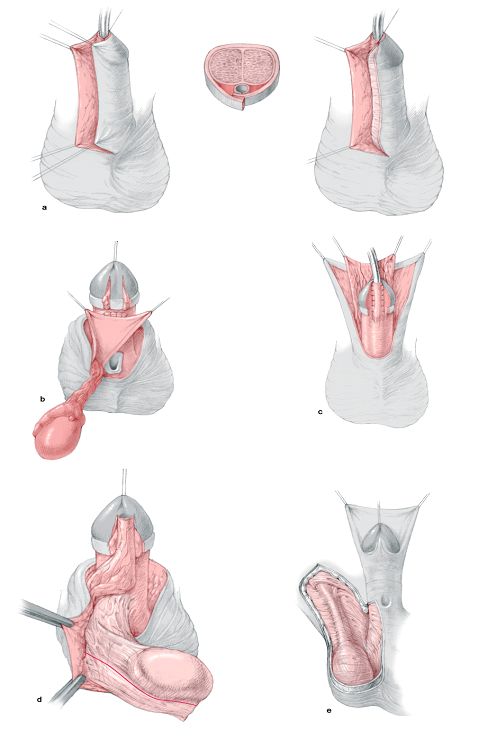
Transverse Preputial Island Flap
Operative steps

A deep Y-shaped incision is made on the glans as in the Lateral based (LB) flap technique. Meticulous excision of any chordee or fibrous bands is carried out. This fibrous tissue is particularly heavy in the midline but may extend well laterally. The meatus is assessed and a cut back is made to widen the meatus. A sub coronal incision is made around the glans. The incision continues laterally until it reaches the gap where the fibrous chordee was excised.
The penile and preputial skin is dissected free off the shaft from distal to proximal close to the Buck's fascia preserving the arteries that constitute the pedicle to the preputial flap.
A 1.5 cm wide rectangular flap is prepared. The length must suffice the gap between the meatus and the tip of the glans. Extra length can be obtained by going down into the penile skin in a horseshoe fashion on either side. The flap is tubularised around a 10 Fr catheter and sutured into the meatus beginning with the suture line underneath the pedicle utilizing interrupted 7-0 polyglactin suture.
Then, the pedicle is separated from the outer preputial skin in a plane just below the intrinsic blood supply of the outer prepuce down to the root of the penis.
The upper small median flap resulting from the Y incision is sutured to the upper dorsal end of the tube. A V is excised from the tip to obtain a slit like meatus. The mobilized glans wings are rotated medially around the neo-urethra. Three transverse mattress sutures maintain firm approximation of the glanular wings in the midline. The mobilized glans wings are rotated medially and three transverse mattress sutures maintain firm approximation of the glanular wings in the midline. De-epithelialisation of skin to protect the neourethra.

Complications
Fistula, wound disruption, diverticulum and rotation occur in 10 - 30 % of patients.

 
MAGPI (Meatal Advancement and Glanuloplasty Incorporated)

This technique may be used in glanular hypospadias with mobile urethral meatus that can be pushed to the tip of the glans. If the meatus is not mobile enough, the results are less satisfactory.
Meatal advancement: The dorsal lip distal to the meatus is cut longitudinally to avoid urine deflecting downwards. In the classic MAGPI, the incision is closed transversely (Heineke Mickulicz technique). Thus the dorsal meatal edge is advanced distally. Recently, some surgeons leave it without closure as a modification from Snodgrass technique.
The glanuloplasty is accomplished by elevating the ventral edge of the meatus forwards and rotating the flattened glanular wings upwards and ventrally in a conical manner. It is important to reapproximate glans tissue in a two layers fashion with a deep closure of glans mesenchyme and a superficial layer of glans epithelium. There have been several modifications of this technique (Duckett and Baskin, 1996).

Complications
Meatal regression may occur if the technique is used in patients with immobile urethral meatus. Precision is required to achieve a conical glans.


Onlay Island Flap

The Onlay Island Flap is ideal for patients with proximal hypospadias without deep Chordee. According to the author experience, most patients with proximal hypospadias have deep chordee that necessitates excision. However, recently, many surgeons prefer to perform dorsal placation if the chordee is less than 30o after skin degloving and preserve the urethral plate.

Operative steps
The tip of the neo-meatus is identified. This point is where the flat ventral surface of the glans begins to curve around the meatus. A midline vertical incision is made in the glans until the width of the glanular groove is adequate for the meatus. The vertical incision is left open without closure for secondary epithelialisation.
A subcoronal incision is made around the glans. The incision continues on either side of the urethral plate at the junction with the normal ventral skin, then up on either side of the glanular groove to the apex of the glansplasty.
The skin is degloved from distal to proximal close to the Buck's fascia preserving the arteries that constitute the pedicle to the preputial flap. The pedicle is then separated from the outer preputial skin in a plane just below the intrinsic blood supply of the outer prepuce. The elevation of the glans wings will permit them to be rotated around the urethroplasty.
A 1-cm wide onlay flap is prepared from the inner prepuce. The onlay flap is sutured into place beginning with the suture line underneath the pedicle utilizing running 7-0 polyglactin suture. The glans should be drawn together setting up the first stitch of the glansplasty ventrally at its apex.
The mobilized glans wings are rotated medially around the neo-urethra. Three transverse mattress sutures maintain firm approximation of the glanular wings in the midline.

Complications
Fistula, wound disruption, rotation, recurrent curvature occurs in 10 - 20 % of patients.


Two Stage repair

A small group of patients with severe proximal hypospadias, chordee, and a small phallus as well as patients with recurrent hypospadias and fibrous unhealthy skin may benefit from a two-stage procedure (Fig. 10).
In the first stage, a circumferential incision is made proximal to the coronal sulcus, the chordee is excised, and the penile shaft is de-golved. Penile straightening and removal of all chordee tissue must be confirmed by the use of the artificial erection test.

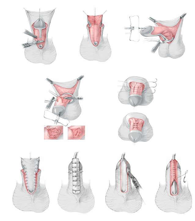
Fig. 10: Steps of two stage repair: identification of chordee, excision of ventral chordee and plication if needed. Coverage of raw surface with skin graft. Tubularisation as a final step

The glans is divided deeply in the midline to the tip. The dorsal foreskin is unfolded carefully and divided in the midline. A midline closure is performed, and the midline sutures catch a small portion of Buck's fascia. The bladder is drained with an 8 French Silastic Foley catheter for approximately 5 to 7 days.
If there is inadequate genital skin available, buccal mucosa or rarely bladder mucosa may be used. The buccal mucosa is harvested from the inner surface of the cheek or the inner surface of the upper or lower lip. The parotid duct is identified opposite the upper molars, and cannulated with 3-0 nylon. The graft is outlined and the submucosa infiltrated with 1% lignocaine containing 1:2000 epinephrine. The graft is incised and the mucosa is dissected away by sharp dissection.
The second stage of the procedure is carried out 6 to 12 months later. The previously transferred skin or mucosa is used to reconstruct the glans and urethra. A 16-mm diameter strip is measured, extending to the tip of the glans. The strip is tubularized with a running subcuticular stitch of 6-0 Vicryl® all the way to the tip of the glans. Tension is reduced by generous mobilization and undermining of adjacent tissues. A protective intermediate layer (either tunica vaginalis or dartos) helps to reduce post-operative complications.
The lateral skin edges are mobilized, and the remaining tissue is closed over the repair in at least two layers. A strip of skin (3 - 5 mm wide) is de-epithelialised on one side to provide a raw surface of deep dermis. This is achieved by cutting 2 or 3 fine longitudinal strips with a pair of small curved-on-scissors. The medial edge of the shaved flap is brought across the buried urethroplasty and sutured to fascial tissue beneath the other flap (double breasting).


Artificial erection test and chordee (curvature) correction

Ventral curvature (chordee) may be evaluated by the artificial erection test. There are two types of chordee associated with hypospadias: 1) Chordee associated with distal hypospadias (skin chordee). This superficial chordee is subcutaneous, proximal to the meatus and can be corrected by mobilization of the skin proximal to the meatus. 2) The other type of chordee is commonly associated with proximal hypospadias. It is usually deep, fibrous and located distal to the meatus. This curvature may be corrected either by Heineke Mikulicz technique, dorsal placation, corporal rotation or the "Split & Roll technique".


Use of protective intermediate layer (Fig. 11)

The use of an intermediate or interposition layer between the neourethra and the skin layer has greatly improved the results following hypospadias surgery and reduced complications. Types of protective intermediate layer include:

a) Durham Smith (1973) de-epithelialization
b) Snow (1986) described the use of Tunica vaginalis wrap.
c) Retik (1988) was the first to use dorsal subcutaneous flap from the prepuce.
d) Motiwala (1993) described the use of Dartos flap from the scrotum.
e) Yamataka (1998) reported the use of external spermatic fascia flap.

Fig. 11: Methods for protective intermediate layer
 

Some technical points

1) Stenting: several studies showed that stenting of the neourethra may be associated with more      complications.

2) Dressing: several studies also showed that the type of dressing has a major impact on the      outcome of surgery, some studies showed better results without any dressing at all.

3) In general, if a complication occurs, one should not operate again before 6 months to allow time      for complete healing of the tissues and to give better chance for the success of surgery.

4) Fistula management: The most important two steps in the management of fistula are to exclude      distal obstruction and to excise the cornu of the fistula to reduce the chances of recurrence.

5) Failed distal hypospadias repair: the technique to be adopted depends on the degree of fibrosis
     and the amount of healthy tissue available. Failed Mathieu repair does not necessarily mean that
     we can not do another Mathieu repair.

6) Failed proximal hypospadias: the most important step is to excise all the unhealthy tissues. Then,      according to the healthy tissues available, one may use lateral based flap, a graft … etc.

7) If scrotal transposition is present, it is suggested to correct hypospadias first and then correct the
     scrotal transposition at a later stage to ensure adequate blood supply to the flap used in      urethroplasty.

8) The role of tissue culture and tissue engineering is a point of major research. So far the      indications for tissue culture in the field of hypospadias are still limited.

 
Recommended Reading

Hadidi A, Azmy A (eds.) "Hypospadias Surgery, An illustrated guide" (2004), Springer Verlag, Heidelberg, Germany.


All illustrations and operative descriptions are from the book: "Hypospadias surgery, An illustrated guide", Hadidi A, Azmy A (eds.). All rights are reserved for Springer Verlag, Heidelberg, Germany.