Hypospadias Surgery

by
Professor Ahmed T Hadidi

Professor of Paediatric Surgery, Heidelberg University, Germany
Professor of Paediatric and Plastic Surgery, Cairo University, Egypt


Hypospadias surgery has developed into a well defined art and science. Surgeons dealing with this anomaly should have a detailed understanding of the various basic surgical principles and experience with delicate, precise optically assisted techniques and maintain a clinical workload that is sufficient to obtain consistently good results.

Incidence

One in 300 boys has hypospadias. In the United States a study reported that hypospadias was the most common congenital anomaly among whites. The incidence has been rising during the 1970s and 1980s.
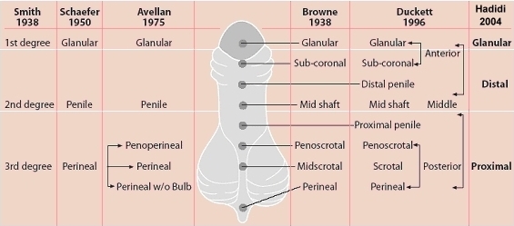
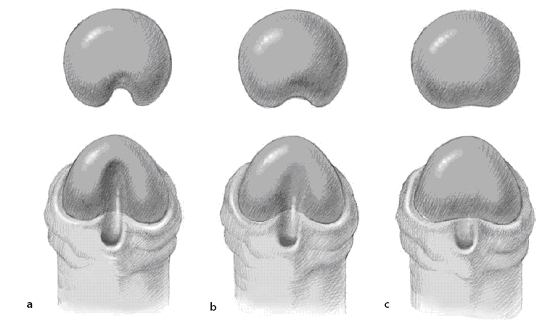
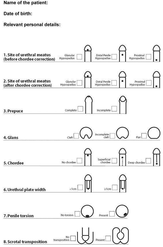
Classification

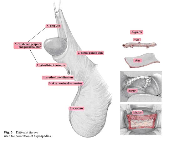
Anatomic classification of hypospadias recognizes the level of the meatus without taking into account curvature. A more recent classification was described. This classification indicates the site of urethral meatus (before and after chordee correction), the prepuce (incomplete or complete), the glans (cleft, incomplete cleft or flat), the width of urethral plate, the degree of penile rotation if present and the presence of scrotal transposition (Fig. 1, 2). Using the general classification (Fig. 4), surgeons are able to conduct multi-centre studies to evaluate different techniques of repair.

Fig. 1: Different classification of hypospadias
Fig. 1: Different classifications of hypospadias, according to location of meatus (modified from Sheldon and Ducket 1987).

Fig. 2 a - c: Classification of glans configuration in hypospadias. (a) Cleft glans. There is a deep groove in the middle of the glans with proper clefting; the urethral plate is narrow and projects to the tip of the glans. (b) Incomplete cleft glans. There is a variable degree of glans split, a shallow glanular groove and a variable degree of urethral plate projection. (c) Flat glans. The urethral plate ends short of the glans penis, no glanular groove. There may be a variable degree of chordee, especially in proximal forms of hypospadias.

 
Timing of Surgery

Recent studies showed that the ideal time for hypospadias correction is between 3 and 15 months as the penis grows less than 1 cm during the first 3 - 4 years (Fig. 3).

Fig. 3: Evaluation of risk for hypospadias repair from birth to age 7 years. The optimal window is from 3 to 15 months of age (modified from Schulz et al. 1983).

Fig. 4: General classification: surgeons are able to conduct multi-centre studies to evaluate different techniques of repair


Different tissues used for correction of hypospadias

Although the penile repairs can be grouped into 8 major principles, depending on the tissues used, each has been subject to countless variations as one surgeon after another adds yet another modification to an already thrice-modified variation of a procedure adapted from a principle derived from the original.
To correct hypospadias and achieve a terminal meatus, one may use one of the following basic principles or tissues: 1) mobilisation of the urethra; 2) skin distal to the meatus; 3) skin proximal to the meatus; 4) preputial skin; 5) combined prepuce and skin proximal the meatus; 6) scrotal skin; 7) dorsal penile skin; 8) different grafts.

1)     Urethral mobilisation

         a)   Urethral mobilisation first described by Beck and Hacker (1897).
         b)   MAGPI described by Duckett (1981, midline vertical incision closed transversely
               and mobilization).
         c)   M configuration by Arap (1984), a modification of MAGPI by placing two sutures on the
               ventral edge.
         d)   UGPI modification of MAGPI by Harrison and Grobelaar (1997) by having a V-shaped
               incision around the original meatus, and having deep glanular wings before urethral
               advancement and upward rotation of the glanular wings.


2)     Skin distal to the meatus

         A)   Use of ventral skin distal to the meatus to reconstruct a completely epithelialized
                 neo-urethra

         a)   U-shaped incision as first described by Thiersch (1869). Notice the U incision is not central
                to avoid suture lines on top of each other.
         b)   Pyramid repair by Duckett and Keating (1989) for Megameatus Intact prepuce (MIP).
         c)   glanular hypospadias with cleft glans.
         d)   DUG repair by Stock and Hanna (1997) combining U-shaped incision with vertical midline
               incision closed transversely.

         B)   Use of ventral skin distal to the meatus to reconstruct a partially epithelialized neo-
                 urethra (Fig. 6)
         a)   Duplay incomplete urethroplasty (1880)
         b)   Denis Browne technique (1949)
         c)   Rich et al (1989) hinging of the urethral plate
         d)   Snodgrass Tubularized Incised Plate (TIP) urethroplasty (1994)

3)     Skin proximal to the meatus

         a)   Wood (1875) described meatal based flap with button hole of prepuce
         b)   Omberdanne (1911) repair, a large round flap, and a purse string suture
         c)   Mathieu repair (1932), a U-shaped incision and two suture lines
         d)   Mustarde repair (1965), a rectangular flap and one suture line
         e)   Barcat balanic groove technique (1969), and a deep midline incision
         f)   Hadidi (1996) Y-V glanuloplasty modified Mathieu. A Y incision in the glans, the center at the               tip of glans, closed as a V and "dog-ears" opened. A small V is excised from the distal end of
              the flap.

4)     Preputial skin

         a)   Button holing of the prepuce described by Thiersch (1869).
         b)   Midline incision of the prepuce described by Edmunds (1913) and Byars (1955).
         c)   Preputial skin as a skin graft to cover the ventral defect of the penis described by Nove-
                Josserand (1897) and Bracka (1995).
         d)   Preputial skin as a free skin graft to form the neo-urethra described by Devine and Horton               (1961).
         e)   Preputial Island Flap as described by Hook (1896), … and Duckett (1980).
         f)   Onlay Island Flap as described by Elder (1987).
         g)   Preputial vascular fascia as a second protective layer described by Retik (1988).

Fig. 6 a - d: Use of ventral skin distal to the meatus to reconstruct a partially epithelialised neourethra: (a) Duplay incomplete urethroplasty (1880); (b) Denis Browne technique (1949); (c) hinging of the urethral plate (Rich et al. 1989); (d) Snodgrass TIP urethroplasty (1994).

5) Combined use of prepuce and the skin proximal to the meatus

         a)   Lateral oblique flap from the side of the penis suggested by Hook (1896).
         b)   One stage repair for proximal hypospadias by Broadbent (1961).
         c)   Parameatal foreskin flap described by Koyanagi (1983).
         d)   Yoke repair described by Snow (1994).
         e)   Lateral based flap combined with Y-V glanuloplasty described by Hadidi (2003).

6) Scrotal skin

         a)   Bouisson (1861) was the first to use scrotal skin for urethral reconstruction.
         b)   Rosenberger (1891) used scrotal tissue for urethroplasty and buried the penis in scrotum.
         c)   Rochet (1899) used a large scrotal flap for total urethroplasty.
         d)   Lowsley and Begg (1938) constructed a long urethral tube from scrotum.
         e)   Beck (1897) suggested Duplay type of urethroplasty and used a rotation flap from scrotum
               for coverage.
         f)   Cecil (1946) used a modification of Rosenberger operation following reconstruction of the
               urethra from ventral penile skin.


7)   Dorsal penile skin

Davis in 1940 tubed the dorsal penile skin with the base proximal in the direction of the circulation. The detached distal end of this tube was passed through a channel in the glans and penis by angulating the penis acutely upward and backward. In the second stage, the proximal pedicle was cut and the penis returned to its normal position. The penile gymnastics required for the Davis procedure apparently seemed too demanding for most surgeons.

8)    Different grafts

         a)   Nove-Josserand (1897) used a split thickness skin graft on a metal probe.
         b)   Devine and Horton (1961) used preputial full thickness skin graft in single stage repair.
         c)   Bracka (1995) used full thickness skin graft in two stage repair.
         d)   Mommelaar (1947) used bladder mucosa for urethral reconstruction.
         e)   Humby (1941) first described the use of buccal mucosa for urethral reconstruction.