| | | | | | | | | | | | | | | | | | | | | | | | | | | | | | | | | | | | | | | | | | | | | | | | | | | | | | | | | | | | | | | | | | | | | | | | | |
| |
|
| |
|
| |
|
General principles | |
|
|
|
|
|
The ideal time for surgery is between 3 and 18 months. The infants are amnesic of the procedure and 70 - 80 % of anomalies can be managed on an outpatient basis.
Fine plastic, micro vascular or ophthalmic instruments including sharp serrated scissors are necessary. Optic magnification is helpful, although low magnification will suffice (X 1-2); some use an operating microscope routinely. Sutures like 6/0 or 7/0 Vicryl (polyglactin 910), Monocryl (poliglecaprone 25) or PDS (polydiaxanone) are used for urethroplasty.
To obtain a bloodless field, a tourniquet (released every 30 - 45 min) or Epinephrine (1:100 000) in 1 % lidocaine is used. Haemostasis should be ensured using bipolar diathermy. Urethroplasty should be performed around a 10 Fr catheter to avoid subsequent stenosis. A compressing dressing is applied post-operatively for 6 hours for hemostasis. The author prefers to remove the dressing after 6 hours but many surgeons prefer to use silastic foam or Tegaderm dressing for 2 - 5 days. The author does not leave a catheter inside the urethra routinely because it causes irritation and interferes with healing. However many surgeons leave a 6 Fr silastic catheter for 7 - 10 days. |
|
|
|
|
|
|
|
|
|
|
|
|
|
|
|
|
|
|
|
|
|
|
|
| |
| |
| |
Choice of operative technique | |
|
|
| |
|
More than three hundred operations have been described for the treatment of hypospadias. Surgeons have proceeded through Browne repairs and scrotal flaps, to Duplay tubes, to free skin grafts, to island flaps and onlays, to bladder and buccal mucosal repairs, to a host of single-stage innovations, to different concepts of chordee correction and with all manner of bladder drainage systems. However, hypospadias repairs can be grouped into five or six major principles, depending on the tissues used.
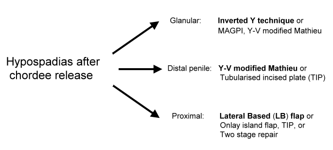
For glanular hypospadias with mobile meatus, the author prefers to use the Inverted Y technique. For distal hypospadias, he prefers to use the Y-V glanuloplasty modified Mathieu approach. The author has adopted the lateral-based flap for proximal hypospadias. Two-stage repair may be preferred in patients with perineal hypospadias to avoid the use of hair-bearing areas of skin. Fig. 7 summarises the author's recommendations for primary hypospadias repair.
|
|
|
|
|
|
|
|
|
|
|
|
|
|
|
|
|
|
|
| |
|
| |
|
|
|
|
|
|
|
|
|
|
|
|
|
|
|
|
|
|
|
|
|
|
|
|
|
|
|
|
|
|
| |
|
Fig. 7: Recommendations for primary hypospadias repair | |
|
|
|
| |
|
| |
|
| |
The Y-V modified Mathieu procedure
The meatal-based flap technique of Mathieu is the most popular technique for distal hypospadias repair and has withstood the test of time. However, the major drawback of the original Mathieu technique is the final appearance of the meatus (a smiling meatus that is not very terminal). The Y-V glanuloplasty helps to employ the Mathieu operation in all forms of distal hypospadias and gives a terminal, slit like meatus. This will include about 70 to 80 % of patients with hypospadias. The only contraindication is the presence of severe chordee distal to the hypospadiac meatus.
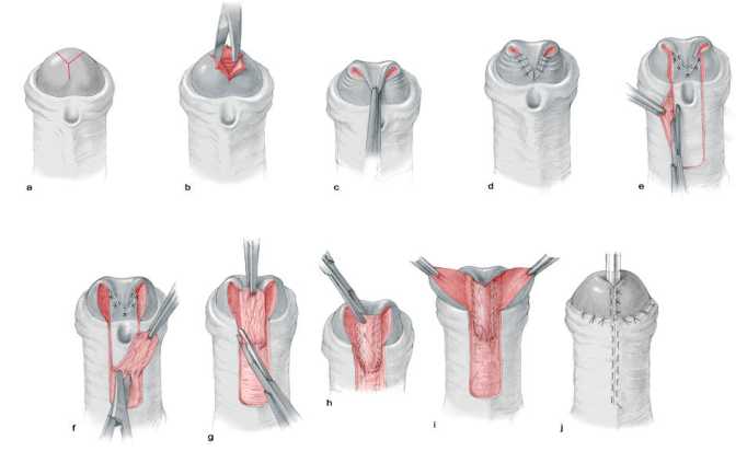
Steps of Y-V glanuloplasty modified Mathieu technique: a) Y Incision; b) The three flaps elevated and coring to make a space for the neo-urethra; c) Y sutured as V with preservation of dog-ears; d) The dog-ears opened; e) U shaped flap; f) urethroplasty; g) A small dog-ear is excised near the meatus; h) A small V is excised from the neourethra; i, ,j) Meatoplasty and glanuloplasty (from Hadidi A; Y-V modified Mathieu in Hadidi A and Azmy A (eds.) Hypospadias Surgery, Art and Science. Heidelberg, Springer Verlag, 2004).
A Y-shaped incision is outlined on the glans with the centre of the Y where the tip of the neo-meatus will be located. Each limb of the Y is 0.5 cm long (Fig. 8 a). The Y-shaped incision is made deep and the three flaps are elevated and a core of soft tissue is excised from the bed of each flap to create a space for the neo-urethra (Fig. 8 b). The Y-shaped gap is sutured as a V making sure to keep the dog- ears at the upper ends of V suture lines (Fig. 8 c). These dog-ears will enlarge the circumference of the tip of glanular wings 1 cm at least (Fig. 8 d).
A U-shaped incision is made slightly longer than the distance between the meatus and the designed tip of the neo-meatus. The longitudinal incisions should diverge away from the hypospadiac meatus to allow for adequate blood supply to the flap. The U-shaped incision will open wide the dog- ears (Fig. 8 e).
A continuous subcuticular running Vicryl® 6-0 on a cutting needle is used for neo-urethra reconstruction. The subcuticular suture is continued until the tip of the glans, then goes back with the same stitch in a running stitch approximating the flap fascia to the depth of the glans and the shaft of the penis (double breasting). Thus, one will have one knot only for the whole two layers (Fig. 8 f).
A small V is excised from the apex of the parameatal flap and the meatus is reconstructed (Fig. 8 g). The glanular wings are approximated using transverse mattress interrupted sutures (Fig 8 h).
The preputial skin is left intact. The prepuce may be reconstructed if parents desire after 6 months when everything has healed well or circumcision performed. Usually, it is not recommended to perform circumcision during urethroplasty, in case complications occur and the preputial skin becomes important.
Complications
Fistula occurs in 2 - 5 % of patients |
|
|
|
|
|
|
|
|
|
|
|
|
|
|
|
|
|
|
|
|
|
|
|
|
|
|
|
|
| |
|
| |
|
|
|
|
|
|
|
|
|
|
|
|
|
|
|
|
|
|
|
|
|
|
|
|
|
|
|
|
|
|
|
|
|
|
|
|
|
|
|
|
|
| |
|
Fig. 8 a - j: Steps of Y-V glanuloplasty modified Mathieu technique. (a) Y incision. (b) Elevation of the three flaps and coring to make a space for the neourethra. (c, d) Y sutured as V with preservation of dog-ears. (e) U-shaped flap. (f) The flap is elevated, taking care to preserve its fascia. (g) The dog-ear is excised from both lateral ends of the flap. (h) Urethroplasty is performed in two layers. (i) A V is excised from the tip of the flap. (j) Meatoplasty and glanuloplasty. |
|
|
|
|
|
|
|
|
|
| |
|
| |
|
| | | | | | | | | | | | | | | | | | | | | | | | | | | | | | | | | | | | | | | | | | | | | | | | | | | | | | | | | | | | | | | | | | | | | | | | | |
| |
Lateral Based (LB) Flap
The lateral based flap may be used in all types of proximal hypospadias This flap with double blood supply, combines the advantages of meatal-based flap, and preputial pedicle flap techniques into one procedure without the need for an intervening anastomosis. It also allows for extensive excision of ventral chordee and the urethral plate (if necessary) without damaging the flap.
Operative steps
A deep Y-shaped incision is made on the glans. The centre of the Y is where the tip of the neo-meatus will be located. The upper two short limbs of the Y are 0.5 cm long. The long vertical limb Y extends down the whole length of the glans penis to the coronary sulcus (Fig. 9 a). The resultant three flaps are elevated and a core of soft tissue is excised to create a space for the neo-urethra (Fig. 9 b).
Meticulous excision of any chordee or fibrous bands is carried out. This fibrous tissue is particularly heavy in the midline but may extend well laterally. The meatus is assessed and a cut back is made to widen the meatus (Fig. 9 c).
A rectangular skin strip is outlined extending proximally from the urethral meatus staying in the midline in the scrotum to avoid potentially hair bearing skin. The skin strip is extended distally and laterally by curving towards the prepuce. This allows for formation of a very long tube that can reach the tip of the glans wherever the original position of hypospadias meatus is (Fig. 9 d).
The skin incision is carried completely around the meatus leaving a small cuff of skin. The meatus is freed proximally. The adjacent penile skin is elevated (rather than the flap). The flap with its pedicle is mobilised through the dorsum of the penis and down to the root of the penis to avoid penile rotation.
The skin strip and proximal cuff are tubularised around a Nelaton catheter size 10 Fr inside the urethra. The author prefers to use Vicryl 6/0 on a cutting needle. Suturing is carried out from distal to proximal in a subcuticular continuous manner. Several reinforcing interrupted stitches are usually taken to form water tight tube (Fig. 9 e).
The neomeatus is then constructed by suturing the terminal end of the neourethra to the central V of the glans. A final slit like meatus is obtained by excising a small V from the tip of the neo-urethra. Then, the glanular wings are wrapped around the neourethra and approximated in the midline. When completed a near normal wide meatus is created at the tip of a conical shaped glans. The long anastomotic contact between the neo-meatus and glans created by the Y glanuloplasty is important to create a wide meatus and avoid post operative meatal stenosis. The vascular areolar subcutaneous tissue layer is then used to provide a complete covering for the neourethra (Fig. 9 f). The skin is closed in the midline using 6/0 Vicryl in a continuous transverse mattress. This helps to simulate the normal ventral median skin raphae (Fig. 9 g). A percutaneous suprapubic cystocath is inserted into the bladder for 10 - 14 days. A compression dressing is applied for 6 hours for haemostasis.
Complications
Fistula occurs in 6 - 12 % of patients. Penile rotation may occur if the pedicle is not mobilized down to the root of the penis. |
|
|
|
|
|
|
|
|
|
|
|
|
|
|
|
|
|
|
|
|
|
|
|
|
|
|
|
|
|
|
| |
|
| |
|
|
|
|
|
|
|
|
|
|
|
|
|
|
|
|
|
|
|
|
|
|
|
|
|
|
|
|
|
|
|
|
|
|
|
|
|
|
|
|
| |
|
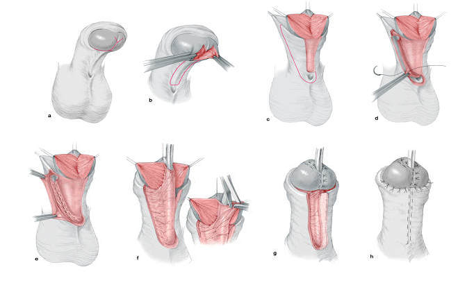
Fig. 9 a - h: Steps of lateral-based (LB) flap technique for single stage repair of proximal hypospadias. (a, b) Y-shaped deep incision of the glans; (c) chordectomy; (d) outline skin incision and flap mobilisation; (e) formation of the neourethra; (f) glanulomeatoplasty; (g) protective intermediate layer; (h) skin closure
|
|
|
|
|
|
|
|
| |
|
|
| |
|
| |
| |
Tubularized Incised Plate Urethroplasty (TIP)
The Tubularized Incised Plate repair (Snodgrass 1994) is based on the assumption that midline incision into the urethral plate may widen it sufficiently for urethroplasty without stricture. Many centres report excellent results with this technique. There are two important criteria to achieve good results: the urethral plate should not be less than 1 cm wide and there should be no distal deep chordee. The technique has gained popularity because it is easily performed, with few complications and results in a slit like meatus. The importance of regular dilatation is still controversial.
Operative steps
A traction suture is placed in the glans just beyond the anticipated dorsal lip of the neomeatus. A circumscribing skin incision is made 1 to 2 mm proximal to the meatus and the shaft skin is degloved to the penoscrotal junction. If a portion of the native urethra is excessively thin, however, a "U" shaped incision is made extending to more healthy tissues.
The urethral plate is separated from the glans wings by parallel incisions along their junction. A tourniquet placed at the base of the penis provides better visualization of the operative field. The glans wings are mobilized avoiding damage to the margins of the urethral plate.
A relaxing incision is made using scissors in the midline from within the meatus to the end of the plate. The incision should not reach the tip of glans. The depth of the relaxing incision depends on the plate width and depth. A 6 Fr stent is secured into the bladder. A 7-0 polyglactin is preferred to tubularize the urethra, with the first stitch placed at approximately the midglans. Tubularization is completed with a 2 layer running subepithelial closure.
Any adjacent dartos tissues are used to cover the neourethra and then a dartos pedicle is developed from the dorsal shaft skin, button-holed, and transposed to the ventrum to additionally cover the repair.
The coronal margins of the glans are approximated with subepithelial 6 - 0 polyglactin. The skin edges of the glans are sutured, and the meatus with 7 - 0 ophthalmic chromic catgut.
Byars' flaps are created from the preputial skin to mimic the median raphe. Subepithelial stitches are used throughout to avoid suture tracks. A tegaderm dressing is applied. The stent is removed approximately 1 week later.
Complications
Fistula occurs in 2 - 15 % of patients. Meatal stenosis 5 - 20 %. |
|
|
|
|
|
|
|
|
|
|
|
|
|
|
|
|
|
|
|
|
|
|
|
|
|
|
|
|
|
|
|
|
|
|
|
|
|
|
|
|
|
|
|
|
|
|
|
| |
|
|
|
 | |
|
|